Glaucoma Surgery

Home | Glaucoma Surgery

This information was adapted from the National Eye Institute to help patients and their families search for general information.
Glaucoma Eye Chart

What is glaucoma?

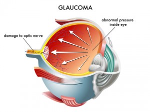
Glaucoma is a group of diseases that can damage the eye’s optic nerve and result in vision loss and blindness. Glaucoma occurs when the normal fluid pressure inside the eyes slowly rises. However, with early treatment, you can often protect your eyes against serious vision loss.

What is the optic nerve?

The optic nerve is a bundle of more than 1 million nerve fibers. It connects the retina to the brain. (See diagram below.) The retina is the light-sensitive tissue at the back of the eye. A healthy optic nerve is necessary for good vision.

What are some other forms of glaucoma?

Open-angle glaucoma is the most common form. Some people have other types of this disease.

  • Low-tension or normal-tension glaucoma. Optic nerve damage and narrowed side vision occur in people with normal eye pressure. Lowering eye pressure at least 30 percent through medicines slows the disease in some people. Glaucoma may worsen in others despite low pressures. A comprehensive medical history is important in identifying other potential risk factors, such as low blood pressure, that contribute to low-tension glaucoma. If no risk factors are identified, the treatment options for low-tension glaucoma are the same as for open-angle glaucoma.
  • Angle-closure glaucoma. The fluid at the front of the eye cannot reach the angle and leave the eye. The angle gets blocked by part of the iris. People with this type of glaucoma have a sudden increase in eye pressure. Symptoms include severe pain and nausea, as well as redness of the eye and blurred vision. If you have these symptoms, you need to seek treatment immediately.
  • Angle-closure Glaucoma. This is a medical emergency. If your doctor is unavailable, go to the nearest hospital or clinic. Without treatment to improve the flow of fluid, the eye can become blind in as few as one or two days. Usually, prompt laser surgery and medicines can clear the blockage and protect sight.
  • Congenital glaucoma. Children are born with a defect in the angle of the eye that slows the normal drainage of fluid. These children usually have obvious symptoms, such as cloudy eyes, sensitivity to light, and excessive tearing. Conventional surgery typically is the suggested treatment, because medicines may have unknown effects on infants and be difficult to administer. Surgery is safe and effective. If surgery is done promptly, these children usually have an excellent chance of having good vision.
  • Secondary glaucomas. These can develop as complications of other medical conditions. These types of glaucomas are sometimes associated with eye surgery or advanced cataracts, eye injuries, certain eye tumors, or uveitis (eye inflammation). Pigmentary glaucoma occurs when pigment from the iris flakes off and blocks the meshwork, slowing fluid drainage. A severe form, called neovascular glaucoma, is linked to diabetes. Corticosteroid drugs used to treat eye inflammations and other diseases can trigger glaucoma in some people. Treatment includes medicines, laser surgery, or conventional surgery.

How does open-angle glaucoma damage the optic nerve?

In the front of the eye is a space called the anterior chamber. A clear fluid flows continuously in and out of the chamber and nourishes nearby tissues. The fluid leaves the chamber at the open angle where the cornea and iris meet. (See diagram below.) When the fluid reaches the angle, it flows through a spongy meshwork, like a drain, and leaves the eye.

Sometimes, when the fluid reaches the angle, it passes too slowly through the meshwork drain. As the fluid builds up, the pressure inside the eye rises to a level that may damage the optic nerve. When the optic nerve is damaged from increased pressure, open-angle glaucoma–and vision loss–may result. That’s why controlling pressure inside the eye is important.

Who is at risk for glaucoma?

Anyone can develop glaucoma. Some people are at higher risk than others. They include:

  • African Americans over age 40.
  • Everyone over age 60, especially Mexican Americans.
  • People with a family history of glaucoma.

A comprehensive dilated eye exam can reveal more risk factors, such as high eye pressure, the thinness of the cornea, and abnormal optic nerve anatomy. In some people with certain combinations of these high-risk factors, medicines in the form of eye drops reduce the risk of developing glaucoma by about half.

Medicare covers an annual comprehensive dilated eye exam for some people at high risk for glaucoma.

What can I do to protect my vision?

Studies have shown that the early detection and treatment of glaucoma before it causes major vision loss, is the best way to control the disease. So, if you fall into one of the high-risk groups for the disease, make sure to have your eyes examined through dilated pupils every two years by an eye care professional.

If you are being treated for glaucoma, be sure to take your glaucoma medicine every day. See your eye care professional regularly.

What are the symptoms of glaucoma?

At first, there are no symptoms. Vision stays normal, and there is no pain.

However, as the disease progresses, a person with glaucoma may notice his or her side vision gradually failing. That is, objects in front may still be seen clearly, but objects to the side may be missed.

As glaucoma remains untreated, people may miss objects to the side and out of the corner of their eye. Without treatment, people with glaucoma will slowly lose their peripheral (side) vision. They seem to be looking through a tunnel. Over time, straight-ahead vision may decrease until no vision remains.

Glaucoma can develop in one or both eyes.
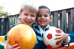
Normal Vision

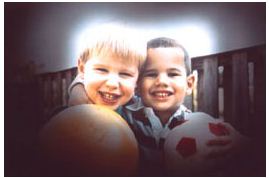
Same scene viewed by a person with glaucoma

How is glaucoma detected?

Glaucoma is detected through a comprehensive eye exam that includes:

  • Visual acuity test. This eye chart test measures how well you see at various distances. A tonometer measures pressure inside the eye to detect glaucoma.
  • Visual field test. This test measures your side (peripheral) vision. It helps your eye care professional tell if you have lost side vision, a sign of glaucoma.
  • Dilated eye exam. Drops are placed in your eyes to widen, or dilate, the pupils. Your eye care professional uses a special magnifying lens to examine your retina and optic nerve for signs of damage and other eye problems. After the exam, your close-up vision may remain blurred for several hours.
  • Tonometry. An instrument (right) measures the pressure inside the eye. Numbing drops may be applied to your eye for this test.
  • Pachymetry. A numbing drop is applied to your eye. Your eye care professional uses an ultrasonic wave instrument to measure the thickness of your cornea.

Can glaucoma be treated?

Yes. Immediate treatment for early-stage, open-angle glaucoma can delay the progression of the disease. That’s why early diagnosis is very important.

Glaucoma treatments include medicines, laser trabeculoplasty, conventional surgery, or a combination of any of these. While these treatments may save remaining vision, they do not improve sight already lost from glaucoma.

  • Medicines. Medicines, in the form of eye drops or pills, are the most common early treatment for glaucoma. Some medicines cause the eye to make less fluid. Others lower pressure by helping fluid drain from the eye.
  • Before you begin glaucoma treatment, tell your eye care professional about other medicines you may be taking. Sometimes the drops can interfere with the way other medicines work.
  • Glaucoma medicines may be taken several times a day. Most people have no problems. However, some medicines can cause headaches or other side effects. For example, drops may cause stinging, burning, and redness in the eyes. Many drugs are available to treat glaucoma. If you have problems with one medicine, tell your eye care professional. Treatment with a different dose or a new drug may be possible.
  • Because glaucoma often has no symptoms, people may be tempted to stop taking or may forget to take, their medicine. You need to use the drops or pills as long as they help control your eye pressure. Regular use is very important. Make sure your eye care professional shows you how to put the drops into your eye. See tips “How should I use my glaucoma eye drops?” on using your glaucoma eye drops.
  • Laser trabeculoplasty. Laser trabeculoplasty helps fluid drain out of the eye. Your doctor may suggest this step at any time. In many cases, you need to keep taking glaucoma drugs after this procedure.
  • Laser trabeculoplasty is performed in your doctor’s office or eye clinic. Before the surgery, numbing drops will be applied to your eye. As you sit facing the laser machine, your doctor will hold a special lens to your eye. A high-intensity beam of light is aimed at the lens and reflected onto the meshwork inside your eye. You may see flashes of bright green or red light. The laser makes several evenly spaced burns that stretch the drainage holes in the meshwork. This allows the fluid to drain better.
  • Like any surgery, laser surgery can cause side effects, such as inflammation. Your doctor may give you some drops to take home for any soreness or inflammation inside the eye. You need to make several follow-up visits to have your eye pressure monitored.
  • If you have glaucoma in both eyes, only one eye will be treated at a time. Laser treatments for each eye will be scheduled several days to several weeks apart.
  • Studies show that laser surgery is very good at reducing pressure in some patients. However, its effects can wear off over time. Your doctor may suggest further treatment.
  • Laser Endocyclophotocoagulation (ECP). ECP is a laser surgery done with or without cataract surgery to lower intraocular pressure. Laser is done inside the eye on the ciliary processes to reduce aqueous production. This treatment can reduce the number of medications used or reduce intraocular pressure (IOP) in some patients. If you are considering cataract surgery ask your surgeon if ECP is a good option for you.
  • Conventional surgery. Conventional surgery makes a new opening for the fluid to leave the eye. (See diagram.) Your doctor may suggest this treatment at any time. Conventional surgery often is done after medicines and laser surgeries have failed to control pressure.
  • Conventional surgery is performed in an eye clinic or hospital. Before the surgery, you will be given medicine to help you relax. Your doctor will make small injections around the eye to numb it. A small piece of tissue is removed to create a new channel for the fluid to drain from the eye. For several weeks after the surgery, you must put drops in the eye to fight infection and inflammation. These drops will be different from those you may have been using before surgery.

As with laser surgery, conventional surgery is performed on one eye at a time. Usually, the operations are four to six weeks apart. Conventional surgery is about 60 to 80 percent effective at lowering eye pressure. If the new drainage opening narrows, a second operation may be needed. Conventional surgery works best if you have not had previous eye surgery, such as a cataract operation.

In some instances, your vision may not be as good as it was before conventional surgery. Conventional surgery can cause side effects, including cataracts, problems with the cornea, and inflammation or infection inside the eye. The buildup of fluid in the back of the eye may cause some patients to see shadows in their vision. If you have any of these problems, tell your doctor so a treatment plan can be developed.

Conventional surgery makes a new opening for the fluid to leave the eye.

How should I use my glaucoma eye drops?

If eye drops have been prescribed for treating your glaucoma, you need to use them properly and as instructed by your eye care professional. Proper use of your glaucoma medication can improve the medicine’s effectiveness and reduce your risk of side effects.